
.jpg)
.png)

.jpg)
.jpg)
-6-25.png)
.png)


-
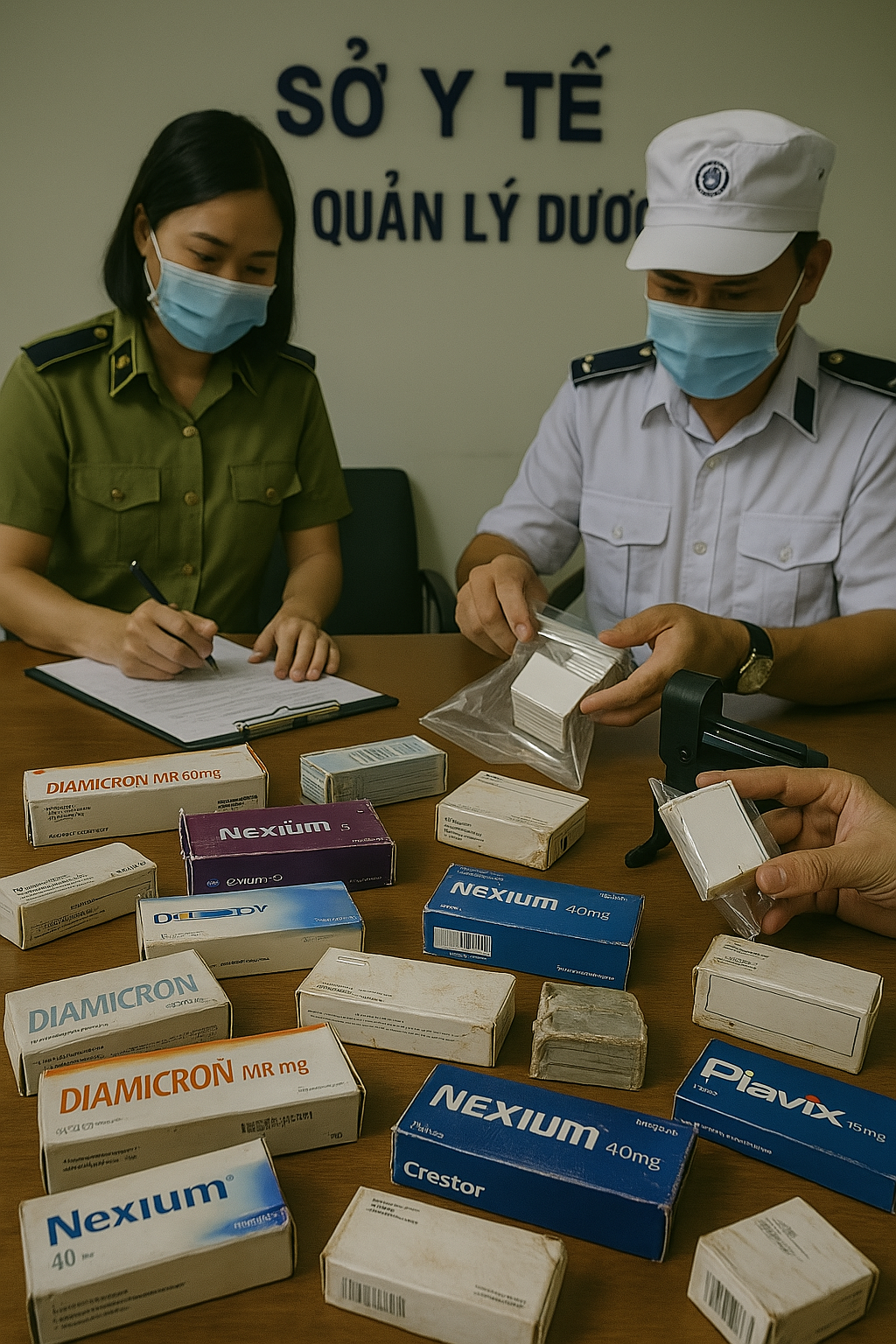
 Tăng cường xử lý tổ chức, cá nhân sản xuất, buôn bán thuốc chữa bệnh và sữa giả
 -
[HƯỚNG DẪN] Đăng ký SmartCA từ ứng dụng VNeID
-
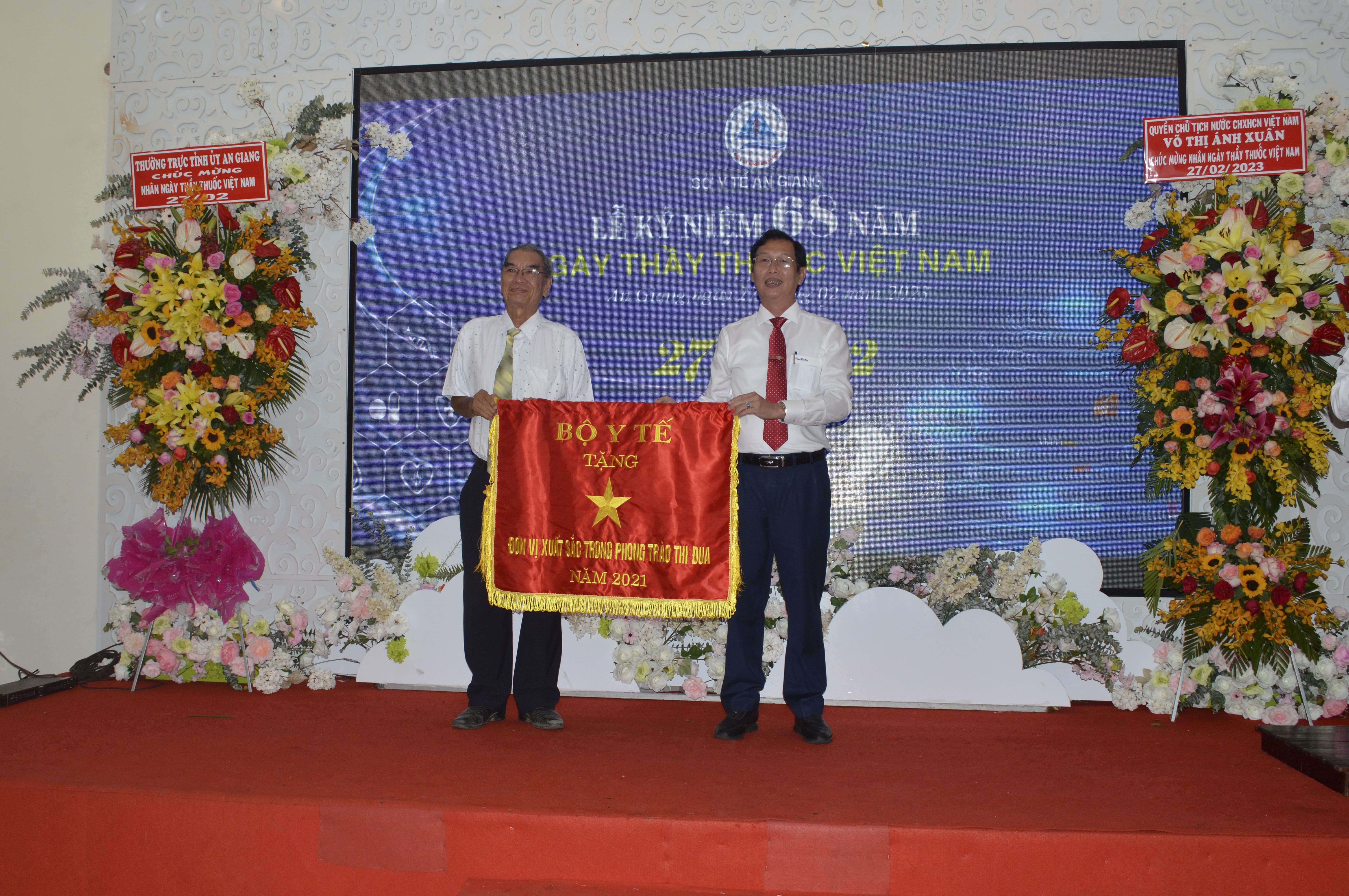
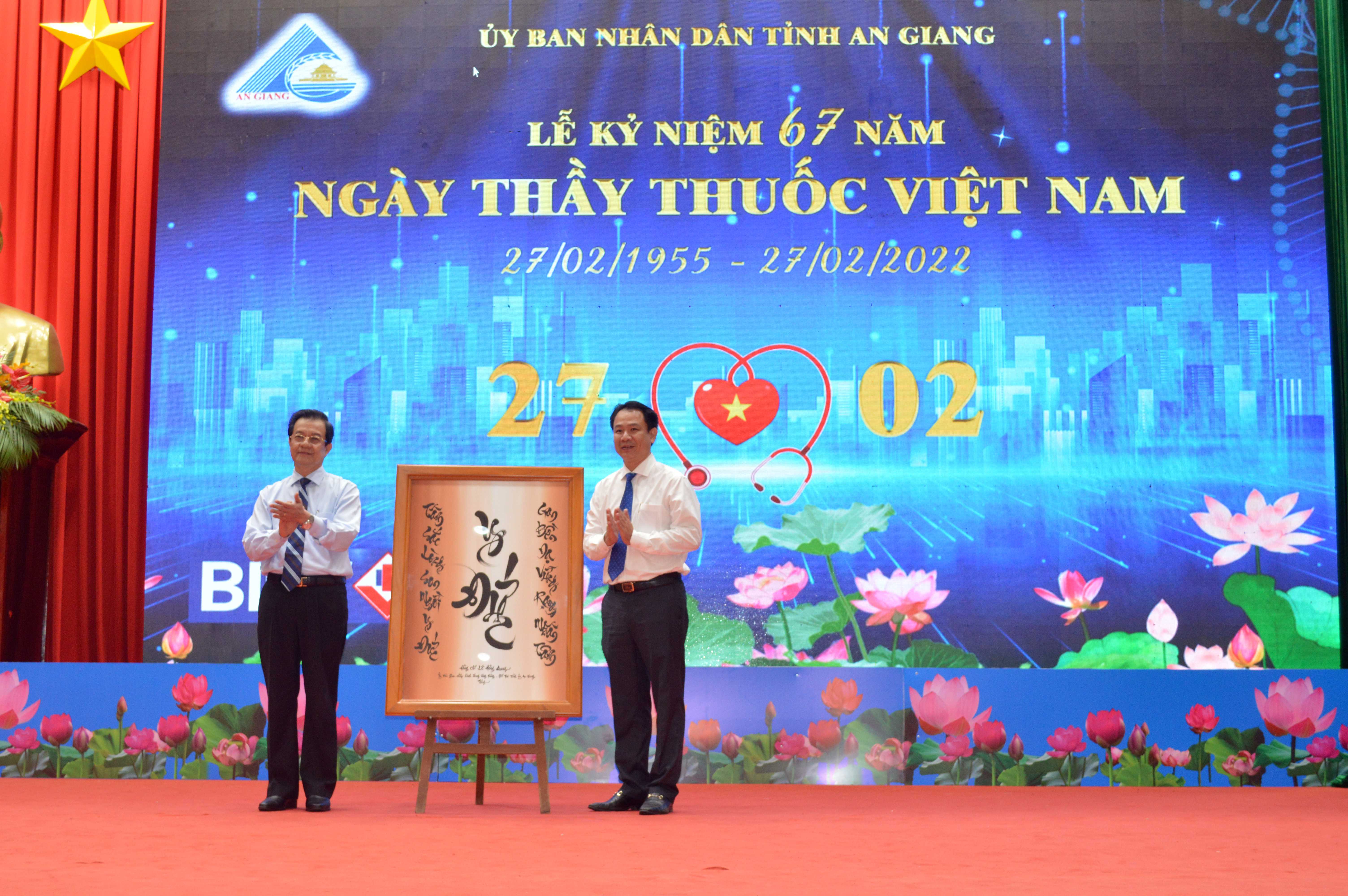
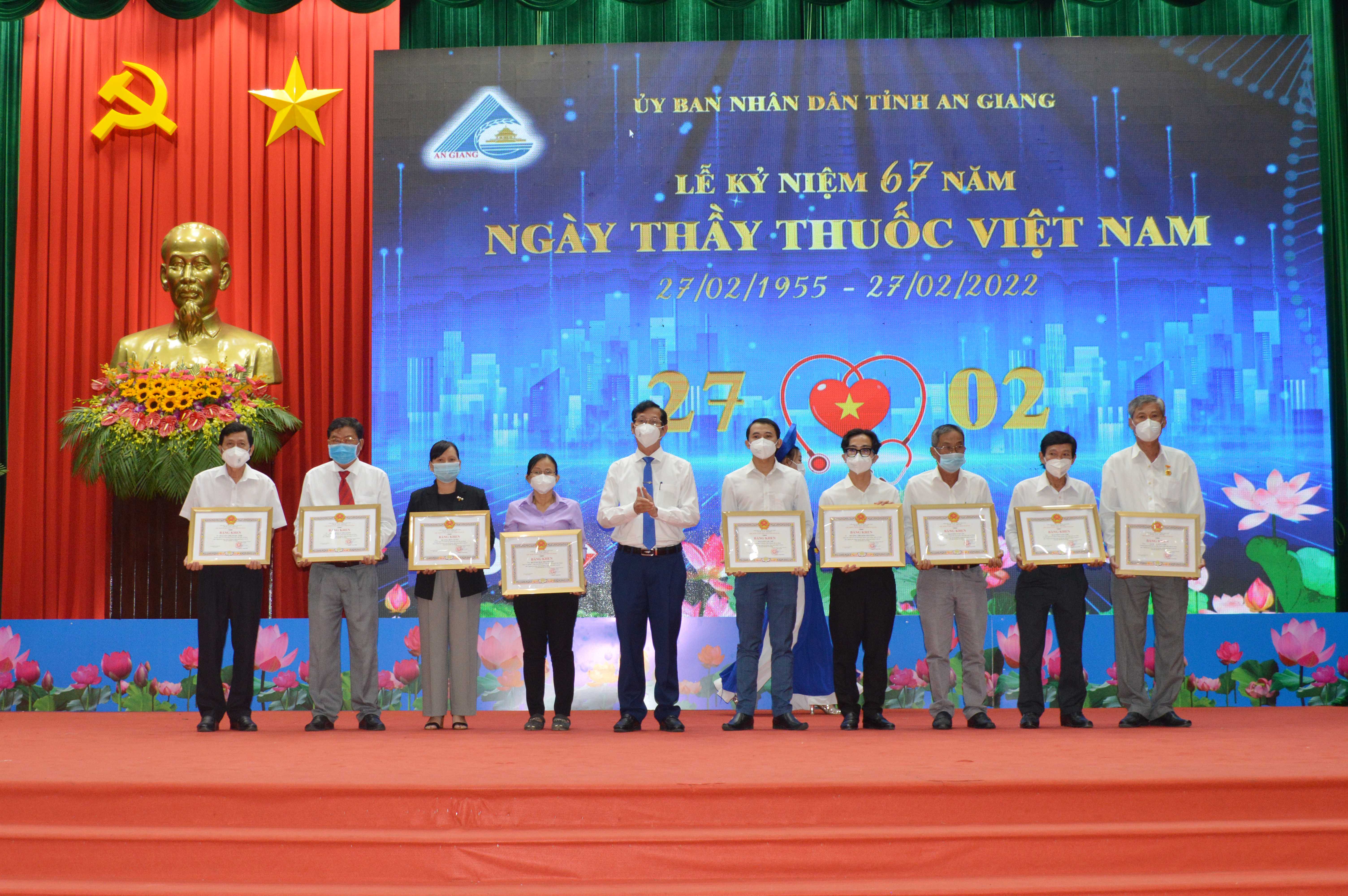
CHƯƠNG TRÌNH LỄ KỶ NIỆM 70 NĂM NGÀY THẦY THUỐC VIỆT NAM - TTVH NT AN GIANG - VĐ NGỌC PHƯƠNG NAM
-
HTV9 | Hội Nghị Đẩy Mạnh Chuyển Đổi Số và Đồng Bộ Dữ Liệu trong Y Tế & Lao Động
-
Dấu ấn công tác y tế 2024: “ĐOÀN KẾT, KỶ CƯƠNG, NÊU GƯƠNG, TRÁCH NHIỆM, HIỆU QUẢ”
-
Sức khỏe và cuộc sống (24-10-2024)
-
Tọa đàm Bệnh lý đột quỵ thực trạng tại An Giang và những tiến bộ trong tiếp cận, điều trị hiện nay
-
TUẦN LỄ THẾ GIỚI NUÔI CON BẰNG SỮA MẸ (1 – 7/8/2024)
-
Thông điệp phòng, chống bệnh bạch hầu
-
Những điểm mới trong Luật Khám bệnh, chữa bệnh (sửa đổi) năm 2023
-
Bệnh viện Đa khoa Y học cổ truyền - Phục hồi chức năng tỉnh An Giang
-
Cách xử trí người bị say nắng, say nóng
-
Khuyến cáo phòng bệnh ho gà
-
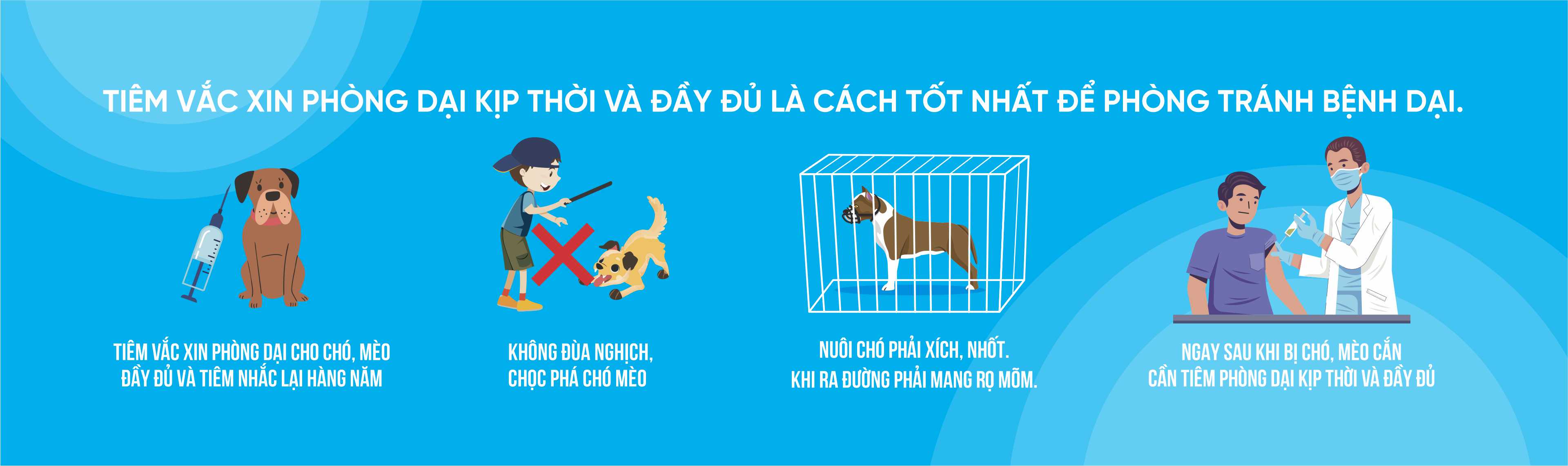
Thông điệp phòng bệnh dại
-
AN GIANG: BỆNH VIỆN Y DƯỢC CỔ TRUYỀN - PHỤC HỒI CHỨC NĂNG CHÍNH THỨC HOẠT ĐỘNG
Cổng thông tin điện tử sở y tế tỉnh an giang